Figure 3 – uploaded by Riia M. Chmielowski

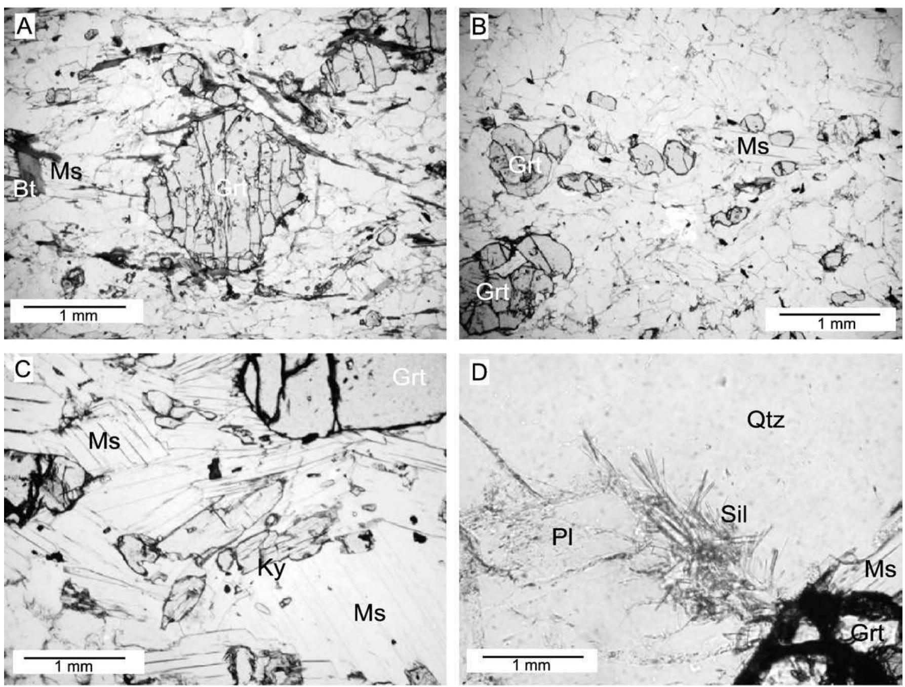
Figure 3 Photomicrographs of microstructures in eclogite from the Collingwood River (Tasmania). Mineral symbols after Kretz (1983) and Phe for phengite. (A) Euhedral porphyroblastic garnet with abundant inclusions in a partly retrogressed matrix. Crossed nicols, sample RCOG11. (B) Fine-grained layer showing textural equilibrium among Grt, Omp. Cam, and Ep. Crossed nicols, sample RO380. (C) Backscattered scanning electron microscope (BSE-SEM) image showing the good textural equilibrium among the mineral phases defining the eclogite facies stage III. Sample RO380. (D) Porphyroblastic garnet and the Di+Pl symplectite after omphacite. Crossed nicols, sample RCO611. (E) BSE-SEM image of a phengite relic surrounded by its destabilization products (Bt +P symplectite). Sample RO380. (F) Anhedral garnet relics, together with abundant porphyroblastic amphiboles. Crossed nicols, sample RO289. (G) BSE image of a porphyroblastic amphibole with a garnet relic as inclusion. Sample RO380. (H) BSE image of a late vein cross-cutting a porphyroblastic amphibole. The vein consists of an association of chlorite, tremolite, and calcite. Sample RO380.
Related Figures (16)


Connect with 287M+ leading minds in your field
Discover breakthrough research and expand your academic network
Join for free
